
 +91-7873-222-000
 WhatsApp

 Book Appointment
Retina Treatment in Delhi: Expert Care for Retinal Detachment, Diabetic Retinopathy & Macular Disease
The retina is the most delicate, vision-critical layer of your eye — and retinal problems often progress silently until vision is at real risk. At Save Sight Centre, Dr. Rajeev Jain, Senior Retina Specialist, provides comprehensive diagnosis and treatment for the full range of retinal and vitreous disorders, from intravitreal injections to complex vitrectomy surgery.
🔎 Quick Answer: Retina Treatment in Delhi
The retina is the light-sensitive layer at the back of the eye, and damage to it — from diabetic retinopathy, retinal detachment, macular degeneration, or vitreous disorders — can cause permanent vision loss if not treated promptly. At Save Sight Centre, a NABH and ISO accredited hospital opposite Adarsh Nagar Metro Station, Dr. Rajeev Jain, Senior Retina Specialist with over 25 years of experience and fellowship training from LV Prasad Eye Institute, Sydney Eye Hospital, and Royal Adelaide Hospital, provides complete retina care — from intravitreal injections (₹15,000 onwards per eye) and laser photocoagulation to scleral buckling and vitrectomy surgery (₹40,000 onwards per eye). Sudden flashes, floaters, or a curtain-like shadow in your vision should be evaluated immediately.
What Do You Need to Know Before Getting Retina Treatment in Delhi?
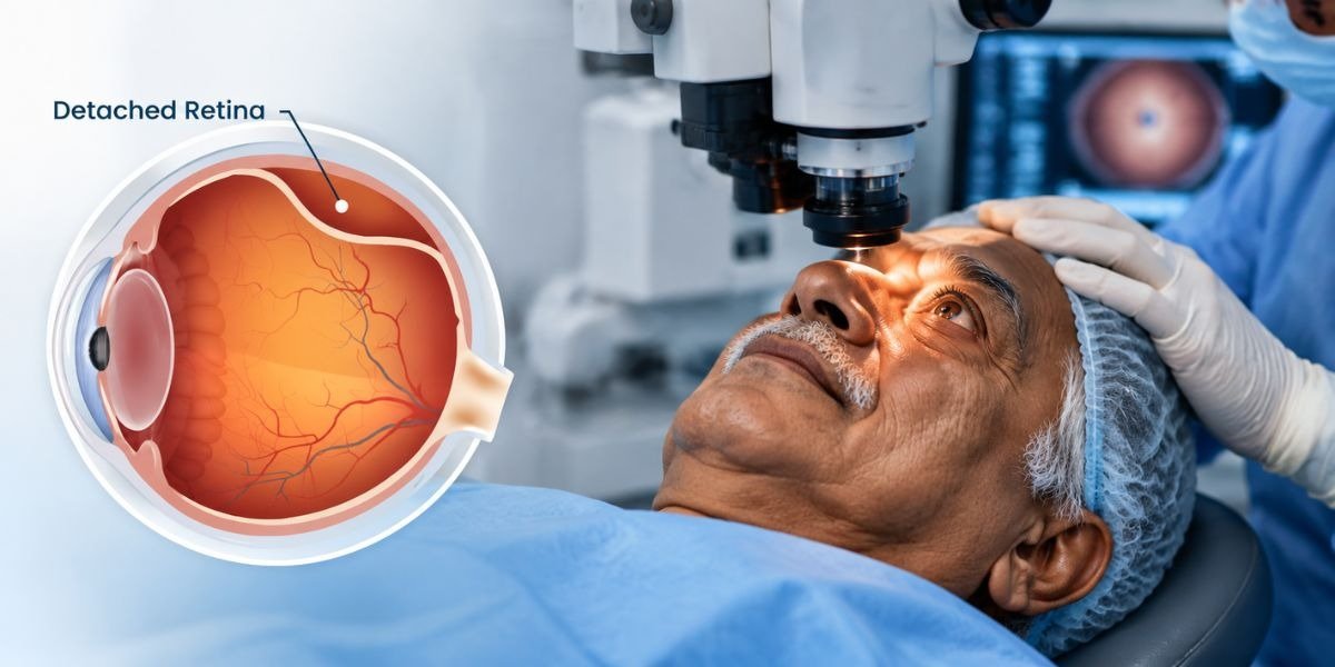
Sudden flashes, floaters, or a shadow/curtain in your vision are warning signs of retinal detachment — a medical emergency requiring urgent care.
Dr. Rajeev Jain, Senior Retina Specialist, has 25+ years of experience and trained at LV Prasad Eye Institute, Sydney Eye Hospital, and Royal Adelaide Hospital.
Treatment ranges from intravitreal injections and laser therapy to scleral buckling and vitrectomy surgery, chosen based on your exact condition.
Diabetics should get a dilated retina exam at least once a year, even without symptoms, since diabetic retinopathy often has no early warning signs.
Consultation and diagnostic test charges (OCT, FFA/ICG, B-scan) apply and are confirmed during your evaluation.
Early diagnosis dramatically improves outcomes — many retinal conditions that would otherwise cause permanent vision loss are treatable when caught in time.
What Is the Retina and Why Does It Need Specialist Care?
The retina is a thin, light-sensitive layer of tissue lining the back of your eye. It works like the film in a camera — capturing light and converting it into electrical signals that travel through the optic nerve to your brain, where they're processed into the images you see. At its centre is the macula, a tiny but critical area responsible for sharp, detailed central vision — the vision you use for reading, driving, and recognising faces.
The vitreous — a clear, gel-like substance that fills the space between the lens and the retina — also plays a role in retinal health. As we age, the vitreous can shrink and pull away from the retina, sometimes causing tears, bleeding, or detachment.
Because the retina has no ability to regenerate once significantly damaged, retinal conditions are treated as time-sensitive. Unlike a cataract, where the lens can simply be replaced, retinal damage that goes untreated for too long can result in permanent, irreversible vision loss — which is why specialist care and prompt diagnosis matter so much.
What Are the Symptoms of Retina Problems?
Many retinal conditions progress silently, while others — like retinal detachment — can develop suddenly and need same-day attention.
⚠ Watch for these warning signs:
- Sudden flashes of light, especially in your side vision
- A sudden increase in floaters — specks, cobwebs, or dark spots drifting across your vision
- A shadow or curtain-like shape blocking part of your vision
- Sudden, painless loss or blurring of vision in one eye
- Distorted vision — straight lines appearing wavy or bent (metamorphopsia)
Who should get a retina check-up regularly?
- Anyone with diabetes, regardless of how well controlled their sugar levels are
- People over 50, especially with a family history of macular degeneration
- Anyone who is highly myopic (high minus power), which raises retinal detachment risk
- People with hypertension or a history of retinal vein occlusion
- Anyone who has had a recent eye injury or blunt trauma
What Are the Most Common Retinal Conditions Treated in Delhi?
Diabetic Retinopathy
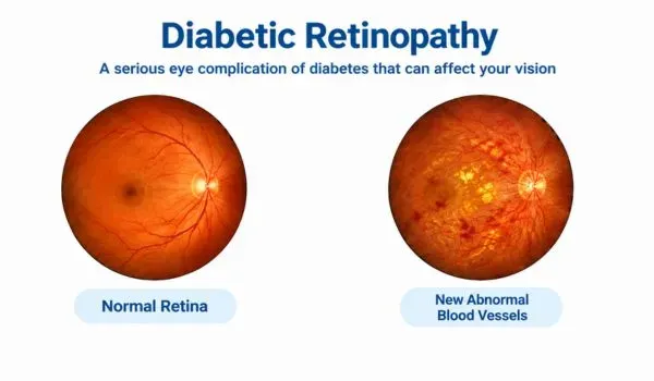
High blood sugar damages the tiny blood vessels in the retina over time, causing leakage, swelling, and abnormal vessel growth that can threaten vision.
Retinal Detachment
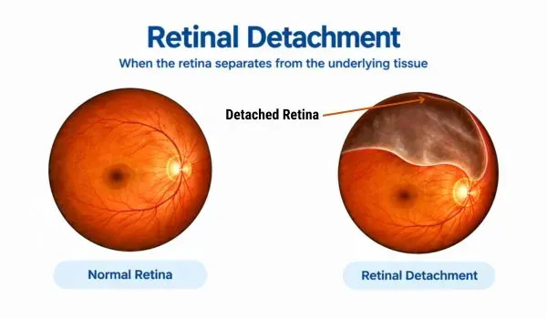
The retina pulls away from the underlying tissue that supports it, cutting off its blood and oxygen supply. Needs urgent surgical treatment to prevent permanent vision loss.
Macular Degeneration (AMD)
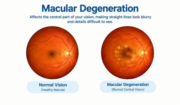
The macula, responsible for sharp central vision, gradually deteriorates with age. Wet AMD progresses faster than dry AMD and needs prompt injection therapy.
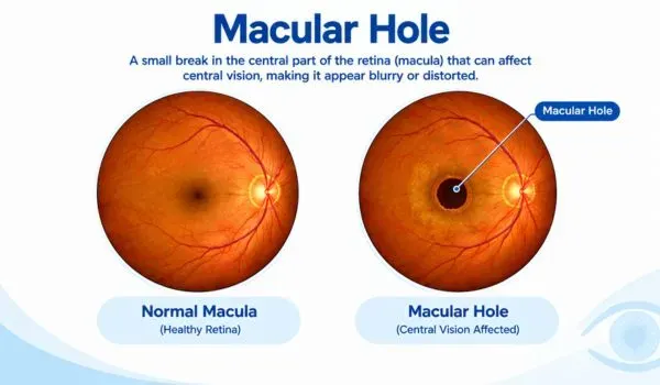
Macular Hole
A small break forms in the macula, usually due to age-related vitreous traction, causing blurred or distorted central vision that typically needs surgical repair.
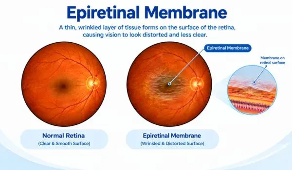
Epiretinal Membrane
A thin layer of scar tissue forms on the retina's surface and can wrinkle or pucker it, distorting central vision. Mild cases are monitored; others need surgery.
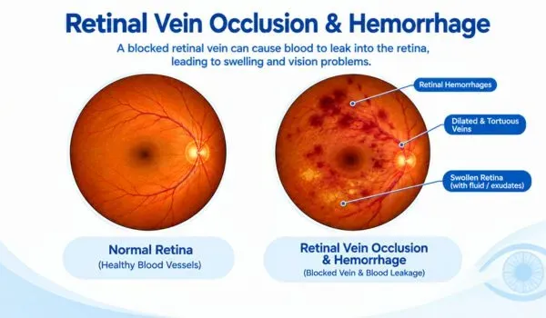
Retinal Vein Occlusion & Hemorrhage
A blocked retinal vein causes blood and fluid to leak into the retina, leading to swelling, hemorrhage, and sudden vision changes. Often linked to hypertension or diabetes.
What Eye Tests Are Used to Diagnose Retina Problems?
Retinal conditions are diagnosed through detailed imaging that lets your specialist see structures at the back of the eye that a routine vision check can't reveal. Our retina workup includes:
Optical Coherence Tomography (OCT)
Captures a detailed cross-sectional image of your retina, detecting macular holes, fluid, swelling, and early degeneration that's invisible on a standard exam. Learn about OCT →
FFA & ICG Angiography
A dye is injected into your bloodstream and photographed as it passes through the retinal blood vessels, revealing leakage, blockages, or abnormal vessel growth. Learn about FFA/ICG →
B-Scan Ultrasound
Used when bleeding or a cataract blocks the direct view of the retina — sound waves create an image of the retina's structure and detect detachment even when it can't be seen directly.
Digital Fundus Photography
High-resolution photographs of the retina document its current condition and are compared over time to track disease progression or treatment response.
Amsler Grid Test
A simple grid test that detects distortion or blind spots in your central vision — an early sign of macular disease that patients can also use at home to self-monitor. Learn about the Amsler Grid →
Dilated Fundus Examination
Your pupils are dilated with drops so your specialist can directly examine the entire retina, including its far periphery, for tears, thinning, or early disease.
How Is Retina Disease Treated in Delhi?
Retina treatment ranges from a simple injection to complex microsurgery, depending on your exact diagnosis. Your retina specialist recommends the least invasive option that will effectively protect your vision.
Non-Surgical Treatment — Many retinal conditions, especially diabetic retinopathy, macular degeneration, and small retinal tears, are effectively managed with injections and laser therapy, without the need for surgery.
Intravitreal Injections (Anti-VEGF / Steroid)
Medication is injected directly into the vitreous cavity to reduce swelling and block abnormal blood vessel growth. Used for diabetic macular edema, wet AMD, and retinal vein occlusion. Starting from ₹15,000 per eye.
Laser Photocoagulation (PRP / Focal)
A laser seals leaking blood vessels or destroys abnormal ones, used for diabetic retinopathy, retinal tears, and lattice degeneration. Cost depends on the extent of laser required — assessed during consultation.
Pneumatic Retinopexy
A gas bubble is injected into the eye to push a detached retina back into place, combined with laser or cryotherapy to seal the tear. Suitable for select smaller, upper retinal detachments. Cost assessed on a case-by-case basis.
Surgical Treatment — Recommended for more complex retinal detachments, large tears, or when non-surgical treatment isn't sufficient to protect vision. Performed by Dr. Rajeev Jain using advanced vitreoretinal surgical techniques.
Scleral Buckling Surgery
A silicone band is placed around the outer eye wall to relieve traction on the retina, helping it reattach. Effective for retinal detachments caused by a retinal tear. Starting from ₹40,000 per eye.
Vitrectomy Surgery
The vitreous gel pulling on the retina is removed, the tear is sealed, and the eye is filled with gas or silicone oil to hold the retina in place. Used for complex or recurrent detachments, macular holes, and vitreous hemorrhage. Starting from ₹40,000 per eye.
Combined Buckle + Vitrectomy
For severe or recurrent detachments, both techniques are combined — the buckle provides external support while vitrectomy removes internal traction. Cost assessed on a case-by-case basis.
What Is Recovery Like After Retina Surgery?
What Should You Avoid After Retina Surgery?
Air Travel with a Gas Bubble
Never fly until your surgeon confirms the gas bubble has fully dissolved — altitude changes can dangerously raise eye pressure.
Heavy Lifting & Straining
Avoid strenuous activity and heavy lifting for at least 2–4 weeks, as straining can raise pressure inside the eye.
Skipping Positioning Instructions
If advised to keep a specific head position, follow it closely — this keeps the gas bubble correctly placed against the healing retina.
Rubbing Your Eyes
Avoid touching or rubbing the operated eye, which can disturb healing tissue and increase infection risk.
Swimming & Water Exposure
Avoid swimming pools and exposing the eye to unclean water for at least 4–6 weeks to prevent infection.
Vitrectomy vs Scleral Buckling: Which Retina Surgery Is Right for You?
Both are effective surgical techniques for retinal detachment, but they work differently and suit different types of cases.
| Basis | Vitrectomy | Scleral Buckling |
|---|---|---|
| How It Works | Internal removal of vitreous traction | External band supports the eye wall |
| Best For | Complex, large, or recurrent detachments | Simple detachments from a single tear |
| Approach | Direct access to the back of the eye | No internal eye surgery required |
| Recovery | May take longer, especially with gas/oil fill | Generally quicker visual recovery |
| Combination | Can be combined with scleral buckle | Can be combined with vitrectomy |
How It Works
Best For
Approach
Recovery
Combination
What Are the Risks of Retina Surgery?
Blurred Vision During Healing
Vision is often blurry in the initial weeks after surgery, especially with a gas bubble in place, and gradually clears as the eye heals.
Cataract Progression
Vitrectomy can accelerate cataract formation, particularly in older patients. This is manageable with standard cataract surgery later if needed.
Elevated Eye Pressure
Gas or oil used to hold the retina in place can occasionally raise eye pressure temporarily, which is monitored and managed with medication.
Re-Detachment
In a small percentage of cases, the retina can detach again, requiring further treatment. Risk varies by the complexity of the original detachment.
Infection or Bleeding
As with any intraocular surgery, infection or bleeding is possible but rare at accredited centres with strict sterile protocols and post-op care.
How Much Does Retina Treatment Cost in Delhi?
Retina treatment cost varies significantly depending on your exact diagnosis and the complexity of the procedure needed. Here's a transparent overview of starting prices at Save Sight Centre:
| Retina Treatment | Starting Price (Per Eye) |
|---|---|
| Intravitreal Injection (Anti-VEGF / Steroid) | ₹15,000 onwards |
| Laser Photocoagulation (PRP / Focal) | On assessment |
| Pneumatic Retinopexy | On assessment |
| Scleral Buckling Surgery | ₹40,000 onwards |
| Vitrectomy Surgery | ₹40,000 onwards |
| Combined Buckle + Vitrectomy | On assessment |
How Much Does Retina Treatment Cost in Delhi?
*Prices shown are starting prices and vary based on your specific diagnosis, complexity, and technique used. Consultation charges and diagnostic test charges (OCT, FFA/ICG, B-scan, etc.) apply separately as advised by your doctor. Confirmed pricing is shared after your evaluation. No-cost EMI is available.
Who Is the Best Retina Specialist in Delhi?

Dr. Rajeev Jain
Senior Retina Specialist with 20+ years of experience treating patients at Save Sight Centre.
Frequently Asked Questions About Retina Treatment in Delhi
Sudden flashes of light, a rapid increase in floaters, or a dark shadow or curtain moving across your field of vision are classic warning signs. These need immediate evaluation — retinal detachment is a medical emergency, and delaying treatment can lead to permanent vision loss.
In select cases of small, simple detachments, pneumatic retinopexy — a gas bubble combined with laser or cryotherapy — can be used instead of open surgery. However, most retinal detachments require scleral buckling, vitrectomy, or both for reliable, lasting reattachment.
No. The eye is fully numbed with anaesthesia before surgery, so you won't feel pain during the procedure. Mild discomfort, grittiness, or watering is common for a few days afterward and is managed with prescribed drops.
Initial recovery typically takes 2–4 weeks, though full visual recovery can take longer, especially if the macula was involved or a gas bubble was used, which can take up to 8 weeks to fully dissolve. Your surgeon will give you a specific timeline based on your case.
Existing damage from diabetic retinopathy generally cannot be fully reversed, but treatment — including injections, laser therapy, and strict blood sugar control — can stop or significantly slow its progression and prevent further vision loss.
Vitrectomy has a high anatomical success rate for reattaching the retina in a single surgery, though outcomes depend on the complexity and duration of the detachment before treatment. Vision recovery varies more than anatomical success, especially if the macula was affected before surgery.
Face-down positioning is sometimes required after vitrectomy for a macular hole, since it helps the gas bubble press against the hole while it heals. Your surgeon will tell you exactly how long and how strictly this is needed for your specific case.
No, air travel is not safe until your surgeon confirms the gas bubble has fully dissolved. Changes in cabin pressure at altitude can cause the bubble to expand rapidly, dangerously raising pressure inside the eye.
Many retina treatments, especially surgical procedures like vitrectomy and scleral buckling for retinal detachment, are often covered under health insurance since they're medically necessary rather than elective. Coverage varies by policy — our team can help you understand what applies to your treatment.
Diabetics should get a comprehensive, dilated retina examination at least once a year, even without any vision symptoms, since diabetic retinopathy often causes no early warning signs. More frequent monitoring may be needed once retinopathy is diagnosed.
Floaters are small specks, cobwebs, or dots that drift across your vision, caused by tiny clumps in the vitreous gel. Flashes are brief streaks or flickers of light, usually caused by the vitreous tugging on the retina. Occasional floaters are common and often harmless, but a sudden increase in either, especially together, needs urgent evaluation.
Macular degeneration currently has no cure, but its progression can be significantly slowed with treatment. Wet AMD is managed with regular anti-VEGF injections, while dry AMD is monitored and managed through lifestyle measures and, in some cases, specific supplements.
This varies widely by condition and individual response — some patients need a short initial course of 3 monthly injections followed by less frequent maintenance doses, while others with chronic conditions need ongoing treatment over years. Your specialist will set a monitoring and treatment schedule based on how your eye responds.
An untreated retinal detachment almost always leads to permanent, irreversible vision loss in the affected eye, as the detached retina is cut off from its blood and oxygen supply. This is why any sudden flashes, floaters, or vision loss should be treated as a medical emergency.
See a retina specialist immediately if you notice sudden flashes, a shower of new floaters, a shadow or curtain in your vision, sudden painless vision loss, or significant distortion of straight lines. These symptoms can indicate retinal detachment or bleeding that needs same-day evaluation.
Don't Delay When It Comes to Your Retina
Book your consultation at Save Sight Centre, Delhi. Our expert team will help you understand your condition, recommend the right treatment and guide you through every step.